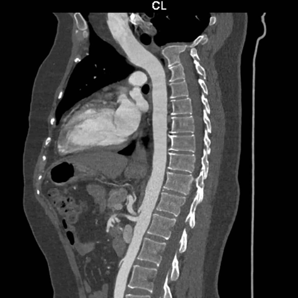
A CT (computed tomography) scan of the cervical spine is a diagnostic imaging procedure that provides detailed cross-sectional images of the cervical (neck) region of the spine. This imaging technique is commonly used to assess and diagnose various conditions affecting the cervical spine. Here are some reasons why a CT cervical spine might be performed and the conditions it can help identify:
- Trauma:
- Evaluation of fractures, dislocations, or other injuries to the cervical spine resulting from trauma, such as car accidents or falls.
- Degenerative Disc Disease:
- Assessment of changes in the intervertebral discs, such as disc herniation, bulging, or degeneration, which may contribute to neck pain or nerve compression.
- Spinal Stenosis:
- Identification of narrowing of the spinal canal or neural foramina, which can lead to compression of the spinal cord or nerve roots.
- Disc Herniation:
- Visualization of herniated discs that may be pressing on spinal nerves, causing pain and neurological symptoms.
- Tumors:
- Detection and characterization of tumors or abnormal growths within the cervical spine.
- Infections:
- Identification of infections affecting the bones or soft tissues of the cervical spine, such as osteomyelitis or discitis.
- Congenital Anomalies:
- Assessment of congenital abnormalities or variations in the structure of the cervical spine.
- Spondylosis:
- Visualization of degenerative changes in the spine, such as osteophytes (bone spurs) or facet joint arthritis.
- Postoperative Evaluation:
- Assessment of the cervical spine following surgical procedures, such as spinal fusion or discectomy.
- Nerve Compression:
- Evaluation of conditions causing compression of spinal nerves, such as foraminal stenosis or radiculopathy.
During a CT cervical spine, the patient lies on a table that moves through the CT scanner, capturing detailed images of the cervical vertebrae, discs, and surrounding structures. The information is processed by a computer to create cross-sectional images.
The interpretation of CT cervical spine images is typically performed by a radiologist, who provides detailed information to guide further medical management. If you have symptoms related to neck pain, neurological symptoms, or have been advised to undergo a CT cervical spine, it’s important to consult with a healthcare professional for a proper evaluation and diagnosis.


