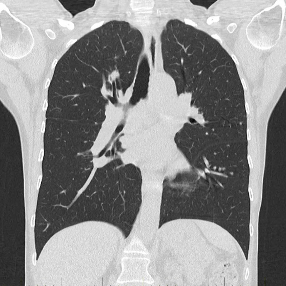
A chest CT (computed tomography) scan is a medical imaging technique that uses X-rays to create detailed cross-sectional images of the chest. It provides more detailed information than a standard X-ray and is commonly used to diagnose and evaluate various conditions affecting the chest, including the lungs, heart, blood vessels, and surrounding structures.
Here are some common reasons why a chest CT might be performed:
- Lung Conditions: To detect and evaluate lung diseases such as pneumonia, lung cancer, pulmonary fibrosis, and bronchiectasis.
- Cardiac Evaluation: To assess the structure and function of the heart, including the coronary arteries. It can be used to identify issues like coronary artery disease, heart valve problems, and pericardial diseases.
- Vascular Conditions: To examine blood vessels in the chest, such as the aorta and pulmonary arteries, to identify aneurysms, dissections, or other vascular abnormalities.
- Infections: To diagnose and assess the extent of infections in the chest, including abscesses or infections in the mediastinum (the area between the lungs).
- Trauma: After chest injuries, a CT scan may be done to assess the extent of damage to the chest structures.
- Tumors: To detect and characterize tumors in the chest, including lung tumors, thymus tumors, and metastases from other parts of the body.
During a chest CT, the patient lies on a table that moves through the CT scanner, which takes a series of X-ray images from different angles. The information is processed by a computer to create detailed cross-sectional images, which can be viewed by a radiologist to make a diagnosis


