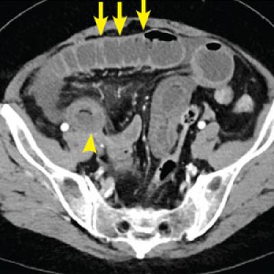
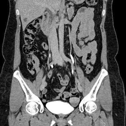
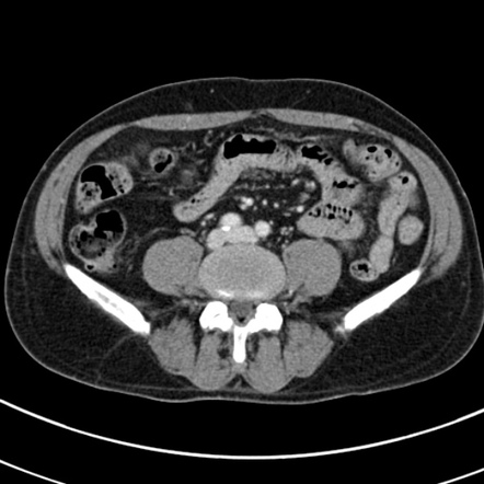
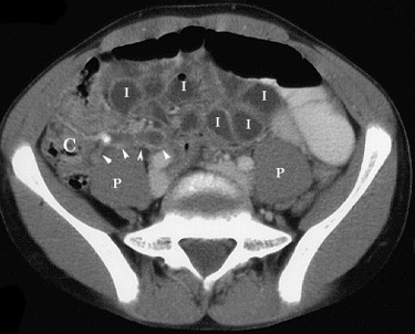
Abdominal pain is a common symptom that can be caused by a variety of underlying conditions affecting the organs within the abdomen. CT (computed tomography) imaging is often used to evaluate the cause of abdominal pain due to its ability to provide detailed cross-sectional images of the abdominal structures. Here are key aspects related to the use of CT in the assessment of abdominal pain:
- Indications for Abdominal CT:
- Unexplained or Severe Pain: Abdominal CT is often recommended when a patient presents with unexplained or severe abdominal pain.
- Trauma: CT may be used to assess for abdominal injuries in cases of trauma, such as motor vehicle accidents or falls.
- Suspected Appendicitis: CT is frequently employed for the evaluation of suspected appendicitis when the clinical presentation is atypical or uncertain.
- Evaluation of Solid Organs: CT is useful for evaluating the liver, spleen, kidneys, and other solid organs for signs of injury, inflammation, or tumors.
- Obstructive Causes: CT can help identify the presence and location of bowel obstructions, which may cause abdominal pain.
- Inflammatory Conditions: CT is valuable for detecting inflammatory conditions such as diverticulitis, inflammatory bowel disease, or pancreatitis.
- Common Findings on Abdominal CT:
- Appendicitis: Enlargement and inflammation of the appendix are visible on CT scans. Periappendiceal fat stranding and fluid accumulation may also be present.
- Kidney Stones: CT is highly sensitive for detecting kidney stones, providing detailed images of the urinary tract.
- Gallstones and Biliary Disorders: CT can reveal gallstones and associated complications, such as inflammation of the gallbladder or bile ducts.
- Abdominal Infections: Inflammatory conditions, such as diverticulitis or intra-abdominal abscesses, may manifest as thickening of the bowel wall or localized fluid collections.
- Tumors and Masses: CT is effective in identifying tumors or masses in various abdominal organs, including the liver, pancreas, and kidneys.
- Bowel Obstruction: Dilated loops of bowel, air-fluid levels, and the site of obstruction can be visualized on CT scans in cases of bowel obstruction.
- Vascular Conditions: CT angiography can be used to evaluate blood vessels in the abdomen, helping identify conditions such as abdominal aortic aneurysm or mesenteric ischemia.
- Contrast-Enhanced CT:
- Intravenous Contrast: The administration of intravenous contrast during the CT scan enhances the visibility of blood vessels and certain organs, aiding in the detection of abnormalities.
- Oral Contrast: In some cases, oral contrast may be used to highlight the gastrointestinal tract, providing better visualization of the stomach and intestines.
- Limitations and Considerations:
- Radiation Exposure: CT involves exposure to ionizing radiation. The decision to perform a CT scan is made based on the clinical situation and the potential benefits of the information obtained.
- Alternative Imaging Modalities: In certain situations, alternative imaging modalities such as ultrasound or MRI may be considered, depending on the clinical scenario and the organs of interest.
The interpretation of abdominal CT scans is typically performed by radiologists, who provide detailed information to guide healthcare professionals in diagnosing the cause of abdominal pain. The decision to perform an abdominal CT scan is made based on the individual’s clinical presentation, medical history, and the need for further diagnostic clarity. If you are experiencing abdominal pain, it is important to consult with a healthcare professional for a thorough evaluation and appropriate management.

