Computed tomography (CT) imaging plays a significant role in the diagnosis and evaluation of tuberculosis (TB), a bacterial infection caused by Mycobacterium tuberculosis. TB primarily affects the lungs, but it can also involve other organs. Here are key aspects related to the use of CT in the context of tuberculosis:
- Pulmonary Tuberculosis:
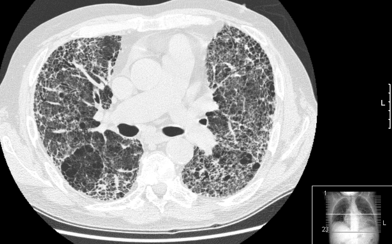
- Parenchymal Involvement: CT is valuable in detecting and characterizing pulmonary involvement in TB. Common findings include nodules, consolidation, and cavities.
- Ghon Focus and Ghon Complex: The Ghon focus represents the primary site of infection, often seen as a small subpleural nodule. A Ghon complex involves the Ghon focus and associated lymphadenopathy.
- Tree-in-Bud Appearance: This appearance is characterized by centrilobular nodules connected by linear structures, representing bronchial inflammation and distal bronchiolar involvement.
- Cavitation: CT is useful for identifying cavities, which are a hallmark of more advanced and active disease.
- Miliary TB: Disseminated TB can present as numerous small nodules throughout the lung, creating a miliary pattern.
- Extra-Pulmonary Tuberculosis:
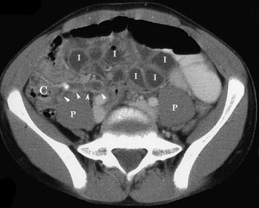
- Lymphadenopathy: CT can visualize enlarged lymph nodes, which are common in extrapulmonary TB. Lymph nodes may exhibit central low attenuation, representing caseation necrosis.
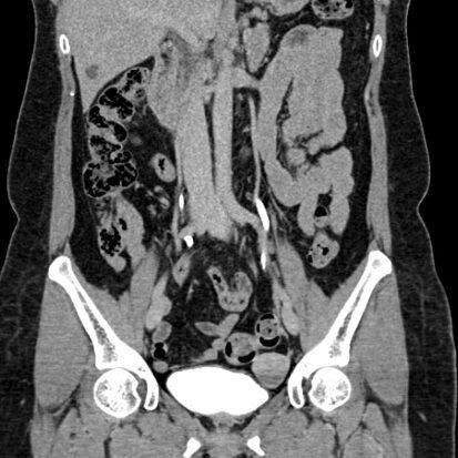
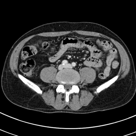
- Abdominal TB: CT is used to assess abdominal involvement, including tuberculous peritonitis, lymphadenopathy, and gastrointestinal TB.
- Skeletal TB: CT helps in evaluating skeletal TB, including involvement of the spine (Pott’s disease), joints, and other bony structures.
- Central Nervous System TB: CT may be employed to assess tuberculous meningitis, hydrocephalus, and intracranial tuberculomas.
- Miliary Tuberculosis:
- Diffuse Involvement: CT is valuable in assessing diffuse involvement of the lungs in miliary TB, with numerous small nodules scattered throughout.
- Follow-up and Treatment Monitoring:
- Monitoring Response to Treatment: CT scans may be used to monitor the response to anti-TB treatment and to assess for complications or disease progression.
- Role in Diagnosis:
- Supplementary Diagnostic Tool: While CT can contribute to the assessment of TB, the definitive diagnosis is typically based on microbiological tests, such as sputum cultures or nucleic acid amplification tests.
- Limitations and Considerations:
- Non-Specific Findings: CT findings in TB can be non-specific and overlap with other infectious or inflammatory conditions.
- Radiation Exposure: Consideration should be given to the potential radiation exposure associated with CT imaging, especially in populations at risk for TB.
- Imaging Guidelines and Protocols:
- Clinical Judgment: The decision to perform a CT scan for TB is based on clinical judgment and guidelines. It is not routinely recommended for all individuals with suspected or confirmed TB.
The interpretation of CT scans in the context of tuberculosis is typically performed by radiologists, and the results are considered alongside clinical and laboratory findings. It’s important to note that CT imaging is just one component of the comprehensive diagnostic approach to tuberculosis. Individuals with suspected TB or those at risk should seek medical attention for appropriate evaluation, diagnosis, and treatment


