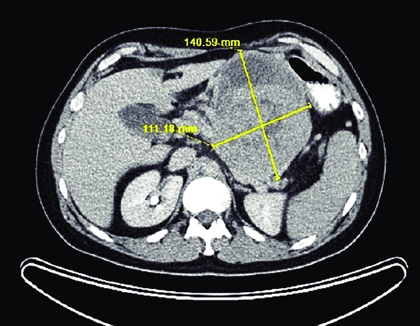
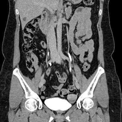
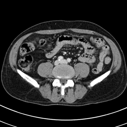
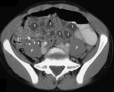
The exploration of the abdomen through the lens of a CT scanner unveils a diverse spectrum of possibilities, ranging from the mundane to the extraordinary. Within this canvas of soft tissues and organs lies the potential for the emergence of neoplasms, or tumors. These abnormal growths, benign or malignant, may reveal their presence in the intricate imagery of a CT abdomen scan.
Let’s embark on a journey through this diagnostic odyssey, where the CT scanner acts as a storyteller, unraveling the tales of abdominal neoplasms:
- Liver Tumors:
- Hepatocellular Carcinoma (HCC): The CT scan may capture focal lesions within the liver, revealing the presence of HCC, often associated with chronic liver disease.
- Hepatic Metastases: Secondary tumors originating from cancers elsewhere may manifest as discreet lesions in the liver.
- Pancreatic Tumors:
- Pancreatic Adenocarcinoma: The CT scan may showcase a mass within the pancreas, often indicative of pancreatic adenocarcinoma.
- Neuroendocrine Tumors (NETs): These tumors, arising from pancreatic islet cells, may present as enhancing lesions on CT.
- Kidney Tumors:
- Renal Cell Carcinoma (RCC): CT imaging can delineate renal masses, aiding in the identification of RCC, the most common kidney cancer.
- Renal Cysts and Oncocytomas: Benign lesions, such as simple cysts or oncocytomas, may also be visualized.
- Adrenal Tumors:
- Adenomas and Adrenocortical Carcinomas: CT can detect adrenal masses, distinguishing between benign adenomas and malignant adrenocortical carcinomas.
- Gastrointestinal Tumors:
- Gastric Cancer: CT may reveal thickening of the gastric wall, suggestive of gastric cancer.
- Colorectal Cancer: Abnormalities in the colonic wall, such as focal thickening or masses, may indicate colorectal tumors.
- Gynecological Tumors:
- Ovarian Cancer: CT imaging can unveil ovarian masses, aiding in the assessment of potential malignancies.
- Uterine Cancer: Uterine masses or thickening of the endometrial lining may be observed on CT.
- Soft Tissue Sarcomas:
- Soft Tissue Tumors: CT may capture soft tissue masses in various abdominal locations, indicating the presence of sarcomas.
- Lymphomas:
- Abdominal Lymphomas: Enlarged lymph nodes and masses within the abdominal lymphatic system may be identified on CT.
- Peritoneal Tumors:
- Peritoneal Carcinomatosis: Disseminated malignancy within the peritoneal cavity may be visualized as enhancing nodules or thickening.
- Neurogenic Tumors:
- Paragangliomas and Neuroblastomas: Tumors arising from neural tissues may be apparent in the retroperitoneum or adrenal glands.
The CT scan, with its cross-sectional prowess, allows for the detection, characterization, and staging of abdominal neoplasms. Radiologists decode the visual narratives, providing clinicians with a roadmap for diagnosis, treatment planning, and prognostic assessment. In the realm of the CT abdomen, each tumor tells a story, and the images bear witness to the intricate tales of pathology and resilience.

