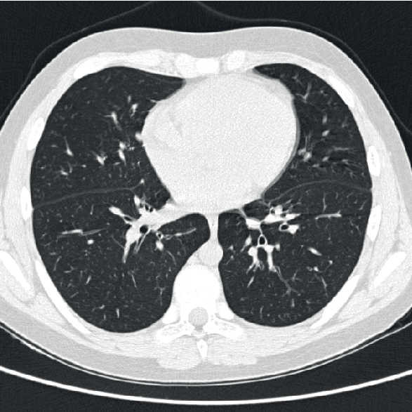
Computed Tomography (CT) of the chest and conventional chest X-rays are both imaging techniques used to assess the thoracic cavity, but they have distinct advantages and applications. Here are some advantages of CT chest over conventional chest X-rays:
- Detailed Anatomical Visualization:
- CT: CT provides detailed cross-sectional images of the chest, allowing for a comprehensive view of the lungs, heart, blood vessels, bones, and other structures. It offers high-resolution images with the ability to visualize fine anatomical details.
- X-ray: Conventional chest X-rays provide a two-dimensional view of the chest, which may lack the detail and clarity compared to the cross-sectional images obtained with CT.
- Increased Sensitivity for Soft Tissues and Lesions:
- CT: CT is highly sensitive for detecting abnormalities in soft tissues, such as lung nodules, tumors, and lymph nodes. It is especially valuable for identifying subtle lesions or abnormalities.
- X-ray: While chest X-rays are useful for detecting certain abnormalities, they may have limitations in visualizing small or subtle lesions, and their sensitivity for soft tissues is lower compared to CT.
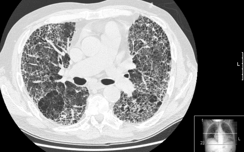
- Evaluation of Lung Parenchyma:
- CT: CT is superior in assessing lung parenchyma, allowing for the detection and characterization of lung diseases such as pneumonia, interstitial lung disease, and pulmonary fibrosis.
- X-ray: Chest X-rays provide a broad overview of lung fields but may have limitations in identifying subtle changes in lung parenchyma.
- Vascular Imaging:
- CT: CT angiography of the chest is valuable for assessing blood vessels, including the pulmonary arteries and veins. It is commonly used to diagnose pulmonary embolism and evaluate vascular abnormalities.
- X-ray: Conventional chest X-rays are limited in their ability to directly visualize blood vessels and are not as effective as CT angiography for vascular assessments.
- Evaluation of Mediastinal Structures:
- CT: CT is excellent for visualizing mediastinal structures, including the heart, thymus, lymph nodes, and major vessels. It provides detailed information about the size, location, and characteristics of mediastinal abnormalities.
- X-ray: While chest X-rays can show an overview of mediastinal structures, CT offers more precise information, making it superior for evaluating mediastinal abnormalities.
- Three-Dimensional Imaging:
- CT: CT provides three-dimensional reconstructions of the chest, which can be valuable for surgical planning and a more comprehensive understanding of complex anatomical relationships.
- X-ray: Conventional chest X-rays are limited to two-dimensional images.
- Quantification of Tissue Density:
- CT: CT allows for the quantification of tissue density, which is useful for distinguishing between different types of tissues (e.g., solid masses, fluid collections) based on their attenuation values.
- X-ray: Chest X-rays provide a grayscale image but lack the quantitative assessment of tissue density offered by CT.
It’s important to note that while CT chest offers numerous advantages, it also involves a higher radiation dose compared to chest X-rays. The choice between the two imaging modalities depends on the clinical question, the level of detail required, and considerations for radiation exposure. In many cases, both imaging modalities may be used complementarily based on the specific diagnostic needs of the patient.