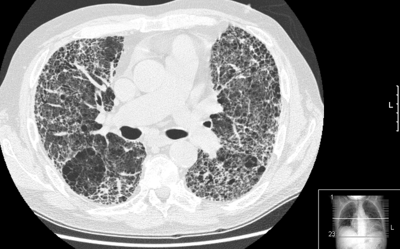
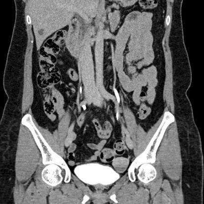
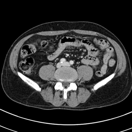
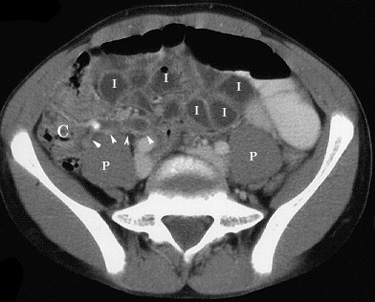
A chest CT (computed tomography) scan is a diagnostic imaging technique that provides detailed cross-sectional images of the chest area, including the lungs, heart, blood vessels, and surrounding structures. It is commonly used to diagnose and evaluate a variety of medical conditions. Here are some common ailments and conditions that can be diagnosed or evaluated using a chest CT:
- Lung Conditions:
- Pneumonia: Inflammation of the lung tissue.
- Lung Cancer: Detection and characterization of lung tumors.
- Pulmonary Nodules: Small, round growths in the lungs that may be benign or malignant.
- Chronic Obstructive Pulmonary Disease (COPD): Emphysema and chronic bronchitis.
- Cardiovascular Conditions:
- Coronary Artery Disease: Visualization of the coronary arteries to assess for blockages.
- Aortic Aneurysm and Dissection: Abnormal enlargement or tearing of the aorta.
- Pericardial Diseases: Inflammation or fluid around the heart.
- Vascular Conditions:
- Pulmonary Embolism: Detection of blood clots in the pulmonary arteries.
- Thoracic Outlet Syndrome: Compression of blood vessels or nerves in the neck and upper chest.
- Aortic Coarctation: Narrowing of the aorta.
- Infections:
- Tuberculosis: Identification of pulmonary tuberculosis.
- Fungal Infections: Assessment of fungal infections affecting the lungs.
- Abscesses: Collection of pus in the lung tissue.
- Interstitial Lung Diseases:
- Idiopathic Pulmonary Fibrosis (IPF): Scarring of the lung tissue with an unknown cause.
- Sarcoidosis: Inflammatory disease affecting multiple organs, including the lungs.
- Chest Trauma:
- Rib Fractures: Assessment of fractures in the ribs.
- Pulmonary Contusion: Bruising of lung tissue after trauma.
- Mediastinal Abnormalities:
- Thymoma: Tumors of the thymus gland.
- Mediastinal Masses: Abnormal growths in the mediastinum (the area between the lungs).
- Airway Conditions:
- Bronchiectasis: Abnormal widening and scarring of the airways.
- Tracheal Stenosis: Narrowing of the trachea.
- Pleural Conditions:
- Pleural Effusion: Accumulation of fluid in the pleural space around the lungs.
- Pleural Thickening: Abnormal thickening of the pleura (lining around the lungs).
The interpretation of chest CT images is typically performed by a radiologist, who analyzes the images and provides detailed information to guide further medical management. If you have symptoms or concerns related to your chest, it’s important to consult with a healthcare professional for a proper evaluation and diagnosis.


